Exercise 31 review & practice sheet: anatomy of the ear presents a comprehensive overview of the intricate structures of the ear and their vital roles in hearing and balance. This exploration delves into the external, middle, and inner ear, unraveling the mechanisms that enable us to perceive sound and maintain equilibrium.
From the sound-gathering auricle to the delicate sensory cells of the cochlea, each component of the ear plays a crucial role in our auditory experience. By embarking on this journey of discovery, we gain a profound appreciation for the remarkable complexity and functionality of the human ear.
External Ear

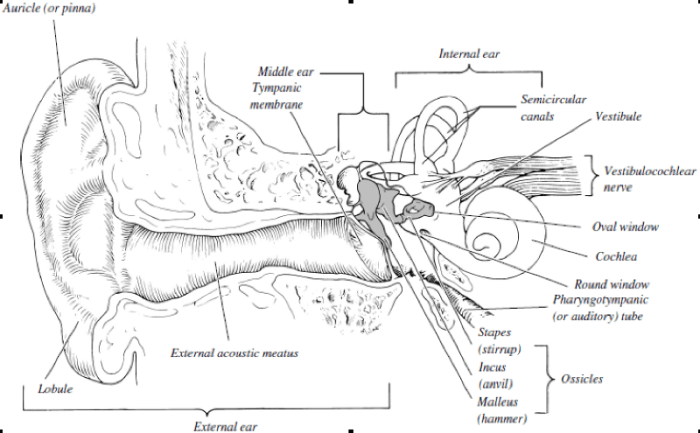
The external ear, also known as the auricle, is the visible part of the ear that collects sound waves and directs them into the ear canal.
Auricle
- The auricle is made up of cartilage covered by skin.
- It has a complex shape that helps to amplify and localize sound waves.
External Auditory Canal
- The external auditory canal is a tube that leads from the auricle to the middle ear.
- It is lined with ceruminous glands that produce earwax, which helps to protect the ear from infection.
Tympanic Membrane
- The tympanic membrane, also known as the eardrum, is a thin membrane that separates the external auditory canal from the middle ear.
- It vibrates in response to sound waves, transmitting them to the middle ear.
Middle Ear

The middle ear is an air-filled cavity that contains three small bones, called ossicles, which transmit sound waves from the tympanic membrane to the inner ear.
Ossicles
- The three ossicles are the malleus, incus, and stapes.
- They are arranged in a chain, with the malleus attached to the tympanic membrane, the incus attached to the malleus, and the stapes attached to the incus.
Eustachian Tube, Exercise 31 review & practice sheet: anatomy of the ear
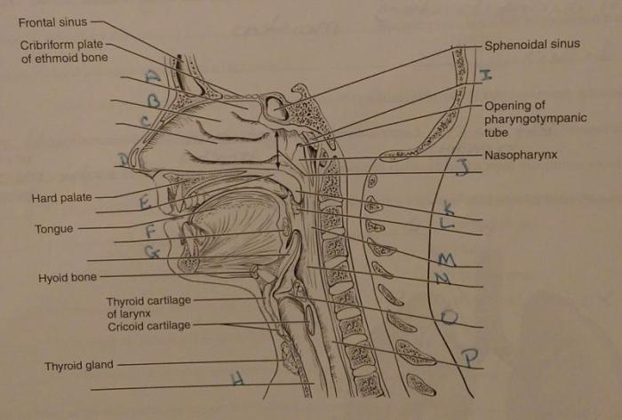
- The Eustachian tube is a narrow tube that connects the middle ear to the back of the throat.
- It helps to equalize air pressure between the middle ear and the outside environment.
Inner Ear
The inner ear is a complex structure that contains the cochlea, semicircular canals, and vestibular apparatus.
Cochlea
- The cochlea is a spiral-shaped tube that is lined with hair cells.
- When sound waves reach the cochlea, they cause the hair cells to vibrate, which generates electrical signals that are sent to the brain.
Semicircular Canals
- The semicircular canals are three fluid-filled tubes that are oriented in different planes.
- They help to maintain balance by detecting changes in head movement.
Vestibular Apparatus
- The vestibular apparatus is a group of structures that help to maintain balance.
- It includes the utricle and saccule, which are two fluid-filled sacs that contain hair cells that detect changes in head position.
Auditory Pathway: Exercise 31 Review & Practice Sheet: Anatomy Of The Ear
The auditory pathway is the neural pathway that transmits sound information from the ear to the brain.
External Ear to Middle Ear
- Sound waves enter the external ear and travel through the external auditory canal.
- They cause the tympanic membrane to vibrate, which in turn causes the ossicles to vibrate.
Middle Ear to Inner Ear
- The vibrations of the ossicles are transmitted to the stapes, which pushes on the oval window of the cochlea.
- This causes the fluid in the cochlea to vibrate, which stimulates the hair cells.
Inner Ear to Brain
- The hair cells in the cochlea generate electrical signals that are transmitted to the auditory nerve.
- The auditory nerve carries the signals to the brain, where they are processed and interpreted as sound.
Hearing Impairments

Hearing impairments can be caused by a variety of factors, including noise exposure, aging, and certain medical conditions.
Types of Hearing Impairments
- Conductive hearing lossis caused by a problem in the outer or middle ear that prevents sound waves from reaching the inner ear.
- Sensorineural hearing lossis caused by damage to the inner ear or auditory nerve.
- Mixed hearing lossis a combination of conductive and sensorineural hearing loss.
Symptoms of Hearing Impairments
- Difficulty hearing faint sounds
- Difficulty understanding speech in noisy environments
- Tinnitus (ringing or buzzing in the ears)
Treatment Options for Hearing Impairments
- Hearing aids
- Cochlear implants
- Surgery
FAQ Overview
What is the function of the tympanic membrane?
The tympanic membrane, commonly known as the eardrum, plays a crucial role in sound transmission. Vibrations in the air are collected by the auricle and channeled through the external auditory canal, causing the tympanic membrane to vibrate. These vibrations are then transmitted to the ossicles of the middle ear, initiating the process of sound transduction.
What is the role of the Eustachian tube?
The Eustachian tube is a vital structure that connects the middle ear to the nasopharynx. It serves two primary functions. Firstly, it equalizes air pressure between the middle ear and the external environment, ensuring optimal sound transmission. Secondly, it facilitates drainage of secretions from the middle ear, preventing fluid buildup and potential infections.
What are the common types of hearing impairments?
Hearing impairments can be classified into two main categories: conductive hearing loss and sensorineural hearing loss. Conductive hearing loss arises from abnormalities in the external or middle ear, such as earwax buildup, otitis media, or damage to the ossicles. Sensorineural hearing loss, on the other hand, results from damage to the inner ear or auditory nerve, often due to aging, noise exposure, or genetic factors.
