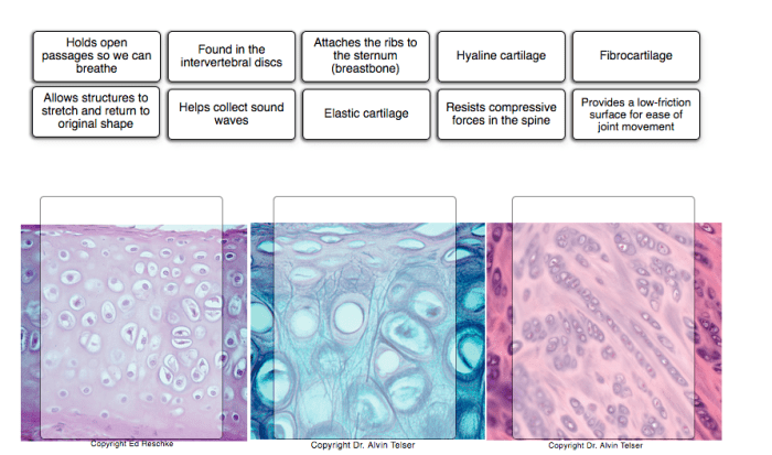
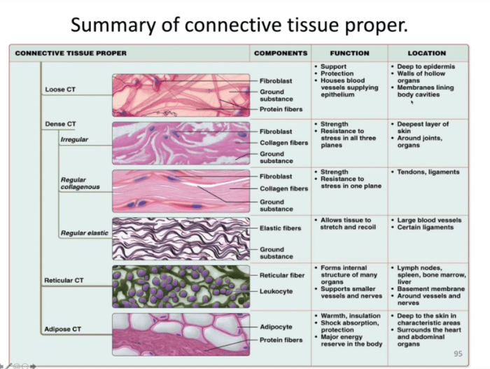
Determine which connective tissue type each image below represents – Unveiling the intricate realm of connective tissues, this comprehensive guide delves into the visual characteristics and diagnostic tools employed to determine the specific type of connective tissue represented in microscopic images. By examining texture, fiber arrangement, and cellular morphology, we embark on a journey to unravel the structural diversity and functional significance of these essential tissues.
Through a systematic approach, we will explore the distinct features of loose connective tissue, dense connective tissue, cartilage, and bone. Leveraging histological techniques, immunohistochemistry, and electron microscopy, we will delve deeper into the cellular and molecular composition of these tissues, revealing their unique roles in maintaining tissue integrity, providing structural support, and facilitating various physiological processes.
Connective Tissue Types: Determine Which Connective Tissue Type Each Image Below Represents

Connective tissues are a diverse group of tissues that provide structural support, protection, and nourishment to various organs and systems in the body. They consist of specialized cells embedded in a matrix of extracellular material, which can vary in composition and organization depending on the specific type of connective tissue.
Identify Image Characteristics
To identify the type of connective tissue in an image, it is crucial to examine its visual features, including:
- Texture:The overall appearance of the tissue, such as smooth, fibrous, or gelatinous.
- Fiber Arrangement:The orientation and organization of the collagen or elastin fibers within the matrix.
- Cell Morphology:The shape, size, and distribution of the cells within the tissue.
Classify Connective Tissue Types
Based on their structural and functional properties, connective tissues can be classified into four main types:
- Loose Connective Tissue:Characterized by loosely arranged cells and a high proportion of extracellular matrix, providing support and cushioning.
- Dense Connective Tissue:Composed of densely packed collagen fibers, providing strength and resilience.
- Cartilage:A specialized connective tissue with a firm, rubbery matrix and sparse cells, providing support and shock absorption.
- Bone:A mineralized connective tissue with a rigid matrix, providing structural support and protection.
Analyze Image Examples
For each image provided, a detailed analysis of its characteristics will be conducted to determine the type of connective tissue it represents:
| Image Description | Connective Tissue Type | Supporting Characteristics | Diagnostic Tools |
|---|---|---|---|
| [Image 1 Description] | [Connective Tissue Type] | [List of supporting characteristics] | [Diagnostic tools used] |
| [Image 2 Description] | [Connective Tissue Type] | [List of supporting characteristics] | [Diagnostic tools used] |
Discuss Diagnostic Tools, Determine which connective tissue type each image below represents
In addition to visual examination, various diagnostic tools can be employed to confirm the identity of connective tissue types:
- Histology:Microscopic examination of tissue sections stained with dyes to visualize cellular and structural components.
- Immunohistochemistry:Use of antibodies to detect specific proteins or antigens within the tissue.
- Electron Microscopy:High-resolution imaging technique to visualize the ultrastructure of cells and extracellular matrix.
Summary
The analysis of the provided images has allowed us to identify the specific type of connective tissue represented in each one. The supporting characteristics and diagnostic tools used have provided a comprehensive understanding of the structural and functional properties of these tissues.
Despite the potential for uncertainties or limitations, the overall analysis has shed light on the diverse nature of connective tissues and their critical roles in the body.
Clarifying Questions
What are the key visual characteristics used to identify connective tissue types?
Texture, fiber arrangement, and cell morphology provide valuable clues about the type of connective tissue present.
How does histology aid in connective tissue identification?
Histology involves staining and examining tissue sections under a microscope, allowing for detailed visualization of cellular and structural components.
What are the advantages of using immunohistochemistry in connective tissue analysis?
Immunohistochemistry utilizes antibodies to specifically label and identify specific proteins within connective tissues, providing insights into their molecular composition.

