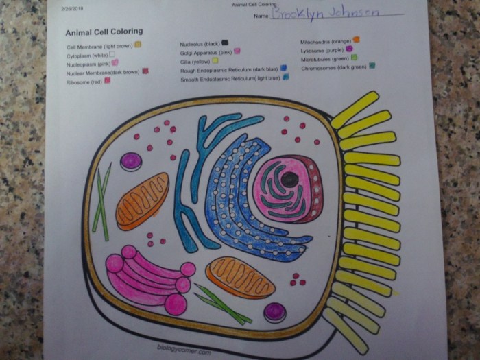
Unveiling the intricate world of animal cells, this guide presents the Animal Cell Coloring Sheet Answer Key, an indispensable tool for students and educators seeking to deepen their understanding of cellular biology. Through vibrant hues and detailed annotations, this answer key illuminates the structures and functions of animal cells, fostering a captivating learning experience.
Delving into the intricacies of animal cells, we explore the significance of coloring as an educational technique, empowering learners to visualize and comprehend complex cellular processes. With step-by-step instructions and comprehensive explanations, this guide empowers educators to effectively incorporate animal cell coloring sheets into their curriculum, unlocking a world of scientific discovery.
Animal Cell Structures
Animal cells are the basic unit of life for all animals. They are composed of a variety of structures, each of which has a specific function. The main structures of an animal cell include the cell membrane, cytoplasm, nucleus, and organelles.
The cell membrane is a thin layer of lipids that surrounds the cell. It protects the cell from its surroundings and regulates the movement of materials into and out of the cell.
The cytoplasm is the jelly-like substance that fills the cell. It contains all of the cell’s organelles, which are small structures that carry out specific functions.
The nucleus is the control center of the cell. It contains the cell’s DNA, which is the genetic material that determines the cell’s characteristics.
Organelles are small structures that carry out specific functions within the cell. Some of the most important organelles include the mitochondria, endoplasmic reticulum, Golgi apparatus, and lysosomes.
| Structure | Function | Image | Notes |
|---|---|---|---|
| Cell membrane | Protects the cell and regulates the movement of materials | [Image of a cell membrane] | The cell membrane is composed of a phospholipid bilayer |
| Cytoplasm | Contains the cell’s organelles | [Image of cytoplasm] | The cytoplasm is composed of a gel-like substance |
| Nucleus | Contains the cell’s DNA | [Image of a nucleus] | The nucleus is surrounded by a nuclear membrane |
| Mitochondria | Produces energy for the cell | [Image of mitochondria] | Mitochondria are often called the “powerhouses of the cell” |
| Endoplasmic reticulum | Folds and transports proteins | [Image of endoplasmic reticulum] | The endoplasmic reticulum is a network of membranes |
| Golgi apparatus | Modifies and packages proteins | [Image of Golgi apparatus] | The Golgi apparatus is a stack of flattened sacs |
| Lysosomes | Digests waste materials | [Image of lysosomes] | Lysosomes are small, membrane-bound vesicles |
Coloring the Animal Cell
Coloring animal cells is an effective educational tool that enhances students’ understanding of cell biology. It allows students to visualize the intricate structures and components of an animal cell, fostering a deeper comprehension of their functions and interactions.Coloring activities engage multiple senses, facilitating memory retention and stimulating visual learning.
They also promote fine motor skills, hand-eye coordination, and attention to detail. Additionally, coloring provides a creative outlet, making the learning process more enjoyable and accessible for students of all ages.
Methods for Coloring Animal Cells, Animal cell coloring sheet answer key
Various methods can be employed for coloring animal cells, each offering unique advantages.
Crayons
Crayons are a traditional and widely available coloring medium. They provide a vibrant and opaque finish, making them suitable for highlighting distinct cell structures. However, crayons may not blend well, resulting in a less refined appearance.
Markers
Markers offer a broader range of colors and can produce a more precise and detailed finish. They are ideal for labeling and outlining specific cell components. However, markers may bleed through paper if applied heavily.
Digital Tools
Digital coloring tools, such as tablets and computer software, provide a versatile and interactive experience. They allow for precise color selection, blending, and zooming, enabling students to create highly accurate and visually appealing representations of animal cells.
Step-by-Step Guide to Coloring an Animal Cell Accurately
To color an animal cell accurately, follow these steps:
- Choose a reliable reference image of an animal cell.
- Identify the different cell structures and their respective colors.
- Use the appropriate coloring medium and apply the colors carefully to each structure.
- Label the cell structures to reinforce their names and functions.
- Add additional details, such as arrows or notes, to highlight specific features or processes.
Answer Key: Animal Cell Coloring Sheet Answer Key
This comprehensive answer key provides the correct colors and additional information for each structure on the animal cell coloring sheet.
By matching the colors and studying the explanations, students can enhance their understanding of the various organelles and their functions within the animal cell.
Nucleus
- Color: Purple
- Description: The control center of the cell, containing the cell’s DNA.
Nucleolus
- Color: Pink
- Description: A dense region within the nucleus where ribosomes are assembled.
Nuclear Membrane
- Color: Light Purple
- Description: A double-layered membrane that surrounds the nucleus.
Cytoplasm
- Color: Light Blue
- Description: The jelly-like substance that fills the cell, containing all the organelles.
Mitochondria
- Color: Red
- Description: Rod-shaped organelles that produce energy for the cell.
Ribosomes
- Color: Green
- Description: Small organelles that assemble proteins.
Endoplasmic Reticulum
- Color: Yellow
- Description: A network of membranes that transports materials throughout the cell.
Golgi Apparatus
- Color: Orange
- Description: A stack of flattened membranes that modifies and packages proteins.
Lysosomes
- Color: Brown
- Description: Sac-like organelles that contain digestive enzymes.
Cell Membrane
- Color: Dark Blue
- Description: A thin layer that surrounds the cell and controls the movement of materials in and out.
Vacuoles
- Color: Light Green
- Description: Membrane-bound sacs that store materials.
Educational Applications

Animal cell coloring sheets are valuable educational tools that can enhance students’ understanding of cell biology and promote scientific literacy. They provide a hands-on and engaging way to explore the structure and function of animal cells.
Learning Objectives
By using animal cell coloring sheets, students can achieve various learning objectives, including:
- Identify and label the major organelles of an animal cell.
- Understand the structure and function of each organelle.
- Visualize the organization and arrangement of organelles within the cell.
- Develop fine motor skills and color recognition.
- Enhance their knowledge of cell biology concepts.
Lesson Plans and Activities
Animal cell coloring sheets can be incorporated into a variety of lesson plans and activities. Some examples include:
- As an introduction to cell biology, to provide students with a visual representation of the different organelles and their functions.
- As a review activity, to reinforce students’ understanding of cell structure and function.
- As a homework assignment, to allow students to practice identifying and labeling organelles at home.
- As part of a project, where students create their own animal cell coloring sheets and present them to the class.
- As a game, where students compete to correctly label and color the organelles of an animal cell.
Variations and Extensions
Animal cell coloring sheets offer numerous variations and extensions that enhance their educational value and cater to diverse learning styles.
Variations
- Organelle-specific coloring sheets:These sheets focus on specific organelles, such as mitochondria, Golgi apparatus, or ribosomes, providing detailed illustrations that facilitate the understanding of their structure and function.
- Cell process coloring sheets:These sheets depict various cellular processes, such as cell division, protein synthesis, or endocytosis, enabling students to visualize and comprehend complex biological mechanisms.
Extensions
- 3D animal cell models:After completing the coloring sheets, students can create 3D models of animal cells using clay, paper-mâché, or other materials. This hands-on activity reinforces their understanding of cell structure and promotes spatial reasoning.
- Research on different cell types:The activity can be extended by assigning students to research different types of cells, such as plant cells, bacterial cells, or specialized animal cells like nerve cells or muscle cells. This encourages them to explore the diversity of cells and their adaptations to specific functions.
These variations and extensions enhance the educational potential of animal cell coloring sheets, making them a valuable tool for fostering a deeper understanding of cell biology.
Answers to Common Questions
What are the benefits of using animal cell coloring sheets in education?
Coloring animal cells enhances visual learning, promotes memorization, and fosters an understanding of cellular structures and functions.
How can I obtain the Animal Cell Coloring Sheet Answer Key?
The answer key is typically provided alongside the coloring sheet or can be accessed through online resources or educational platforms.
What types of animal cells are commonly featured in coloring sheets?
Coloring sheets often depict various types of animal cells, including mammalian cells, plant cells, and specialized cells such as nerve cells or muscle cells.
